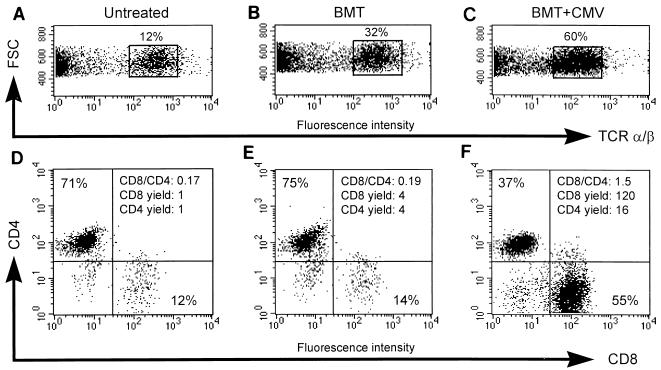
FIG. 3.
Preferential enrichment of CD8 T cells in lung infiltrates. Three-color cytofluorometric analysis of interstitial pulmonary T lymphocytes was done. After perfusion and bronchoalveolar lavage were done for groups of three to five mice, mononuclear leukocytes were recovered from the lung parenchyma by collagenase-DNase digestion and were enriched by Ficoll gradient centrifugation. A lymphocyte gate was set in the forward versus side scatter plot (data not shown). (A and D) Pulmonary lymphocytes recovered as a control from lung tissue of untreated, adult BALB/c mice; (B and E) pulmonary lymphocytes recovered from lung tissue 4 weeks after performance of BMT with 107 syngeneic BMC; (C and F) pulmonary lymphocytes recovered from lung tissue 4 weeks after BMT (same conditions as those described for panels B and E) and concurrent murine CMV infection; (A to C) dot plot of forward scatter (FSC, ordinate) versus PE (TCR α/β, abscissa) fluorescence intensity, with TCR α/β cells enclosed in a rectangle and their percentage among the cells in the lymphocyte gate indicated; (D to F) dot plots of RED613 (CD4, ordinate) versus FITC (CD8, abscissa) fluorescence intensities for TCR α/β-expressing cells. The percentages of CD4 and CD8 T cells are given in the respective quadrants, and the CD8/CD4 ratio is indicated in the upper right quadrant. The yield of CD8 and CD4 T cells is expressed as a multiple of the respective yield from normal, uninfected lungs.

