Fig. 3.

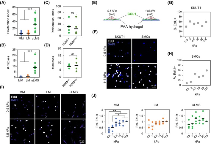
Adhesion to collagen substrates has minimal effect on uLMS cell proliferation. (A) Gene expression‐based proliferation index in normal myometrium (MM; n = 31), leiomyoma (LM; n = 28) and uterine leiomyosarcoma (uLMS; n = 20) tissues indicating enhanced proliferation in uLMS. One‐way ANOVA with post hoc Tukey's test. (B) Number of mitoses per field in MM (n = 7), LM (n = 6) and uLMS (n = 10) tissues. One‐way ANOVA with post hoc Tukey's test. (C, D) Proliferation index (C) and number of mitoses per field (D) in uLMS tumours with high (n = 4) and low (n = 5) high‐density matrix (HDM) indicating no significant differences between groups. Each dot represents one patient tissue; horizontal bars indicate average per group. Two‐tailed Student's t‐test. (E) Schematic representation of the 2D model of controlled collagen substrate stiffness based on polyacrylamide (PAA) hydrogels. (F–H) Representative images (scale bar indicates 50 μm) and quantification of cell proliferation measured by EdU incorporation in SKUT1 cells (n > 95 cells per condition) (G) and primary smooth muscle cells (SMCs; n > 60 cells per condition) (H) at indicated PAA hydrogel stiffness. (I, J) Representative images (I) and quantification (J) of EdU incorporation in MM cells (n = 4 donors), LM (n = 3 donors) and uLMS (n = 4 donors) cells adhered to PAA hydrogel substrates with indicated stiffness. Scale bar indicates 50 μm. Each dot indicates the average per donor; horizontal bars indicate the average per tissue type. One‐way ANOVA with post hoc Tukey's test. ns P > 0.05, *P < 0.05, **P < 0.01, ***P < 0.001.
