Fig. 5.

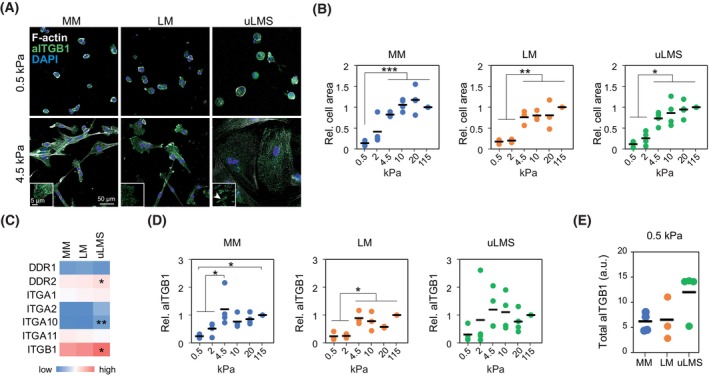
uLMS cells overexpress collagen receptors and show enhanced integrin β1 activity. (A) Representative images of normal myometrium (MM), leiomyoma (LM) and uterine leiomyosarcoma (uLMS) cells adhered to PAA hydrogels showing distinct morphology and presence of focal adhesions at indicated stiffness. At least 50 cells were counted per condition and patient. Scale bar indicates 50 μm. Insets show higher magnification of example cellular edges with elongated focal adhesion in uLMS (arrowhead). Inset Scale bar indicates 5 μm. (B) Quantification of spreading cell area of MM (n = 4 donors), LM (n = 3 donors), uLMS (n = 4 donors) cells on the indicated substrate stiffness. At least 50 cells were counted per condition and patient. (C) Relative collagen receptor gene expression in MM (n = 31), LM (n = 28) and uLMS (n = 17) tissues showing enhanced expression in uLMS of indicated genes. (D) Change in active integrin β1 intensity (aITGB1) at distinct polyacrylamide (PAA) hydrogel stiffness relative to 115 kPa in MM (n = 4 donors), LM (n = 3 donors), uLMS (n = 4 donors) cells. (E) Comparison of total aITGB1 intensity in MM (n = 4 donors), LM (n = 3 donors), and uLMS (n = 4 donors) cells at 0.5 kPa showing a nonsignificant increase in uLMS cells. All statistical analyses were performed by one‐way ANOVA with post hoc Tukey's test. *P < 0.05, **P < 0.01, ***P < 0.001.
