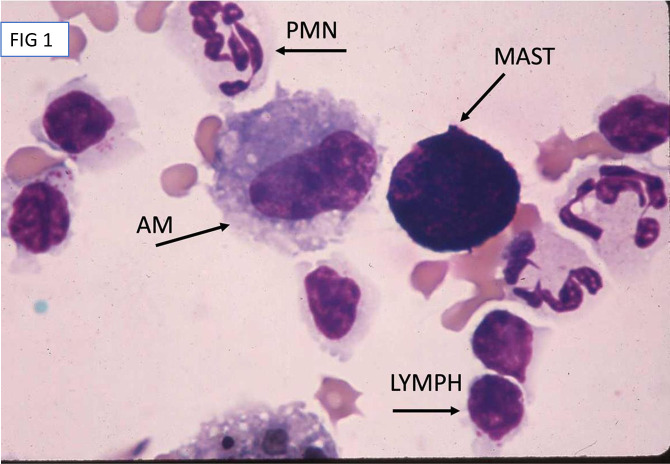
Fig 1. High magnification image of representative cells in BALF.
Sample was prepared via sedimentation, stained with a modified Wright-Giemsa (Diff Quick) and viewed using light microscopy at 1000x under oil. Representatives of cell types are labeled as AM (alveolar macrophage), LYMPH (lymphocyte), PMN (polymorphonuclear neutrophil), and MAST (mast or metachromatic cell).

