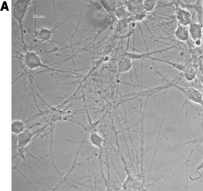
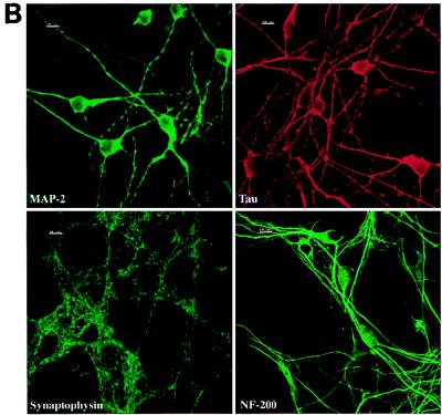
FIG. 1.
Neuron cultures. (A) Phase-contrast microscopy of neurons after 7 DIV. Bar, 10 μm. (B) Staining of the cultures with antibodies specific for neuronal proteins (immunofluorescence, confocal microscopy). The immunostaining for MAP-2, tau, and synaptophysin was done on cultures after 7 DIV, whereas immunostaining for NF-200 was done after 13 DIV. At least 90% of the cells were positive for these markers. Bars, 10 μm.


