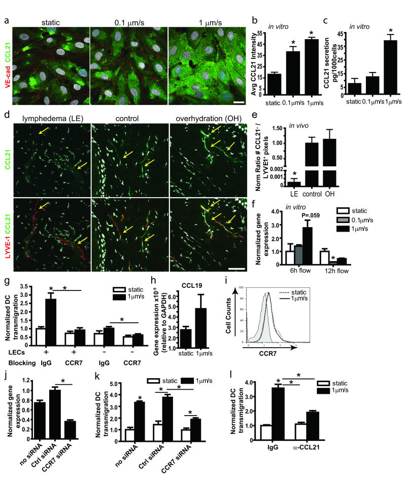
Figure 4. Transmural flow increases CCL21 secretion by lymphatic endothelium.
a, Representative confocal images show CCL21 (green) expressed by lymphatic endothelial cells (LECs) after 12 h exposure to 0 (static), 0.1, and 1μm/sec flow; red, VE-cadherin; bar, 20μm. b, Image quantification of CCL21 protein in cells exposed to 12h flow vs. static conditions. c, CCL21 protein measured by ELISA after 24h culture. d, Representative images of lymphatic vessels (red, LYVE-1) and CCL21 (green) in lymphedematous (LE), control, and overhydrated (OH) skin. Arrows indicate lymphatic vessels; bar, 50μm. e, Quantification of CCL21 staining in cultured LECs after 12h transmural flow. f, Real-time PCR expression after 6h and 12h flow treatment. g, DC transmigration across a LEC monolayer with control IgG or anti-CCR7 blocking antibodies. h, CCL19 gene expression by DCs in 3D cultures after 12h static or flow conditions. i, Representative histogram from flow cytometry showing no differences in CCR7 expression by DCs in static (dotted line) and 12h flow (1μm/sec, solid line) conditions; shaded area shows the negative control. j, Real-time PCR for CCR7 expression in DCs after 24h of siRNA transfection. k, DC transmigration across a LEC monolayer following DC transfection with control or CCR7 siRNA. l, CCL21 blocking inhibits the flow-enhanced DC transmigration across LECs. *P<0.05 compared to static controls.

