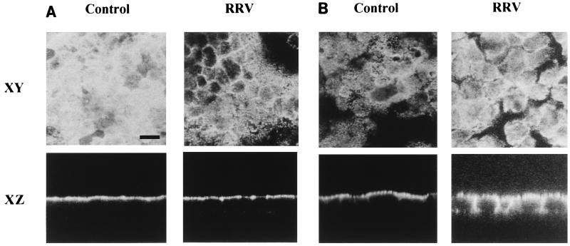
FIG. 2.
Alteration in the apical SI distribution in RRV-infected Caco-2 cells. At 24 h p.i., RRV-infected and mock-infected Caco-2 cells were fixed and permeabilized as described for Fig. 1. (A) SI was stained with a rat anti-SI MAb and fluorescein-coupled anti-rat IgG secondary antibodies. (B) DPP IV was immunostained with a rat anti-DPP IV MAb and fluorescein-coupled anti-rat IgG secondary antibodies. Horizontal (XY) sections at the apical level and vertical (XZ) sections were obtained by direct confocal analysis. Each bar indicates 10 μm.

