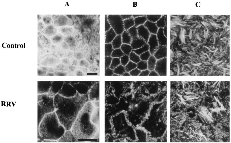
FIG. 7.
Three-dimensional F-actin alteration induced by RRV infection. At 24 h p.i., RRV-infected and mock-infected Caco-2 cells were fixed, permeabilized, and stained with fluorescein-phalloidin, which binds to F-actin. Horizontal sections were generated by CLSM along an axis perpendicular to the monolayer, at the apex (A), the middle (B), and the base (C) of the cell. Bars, 10 μm.

