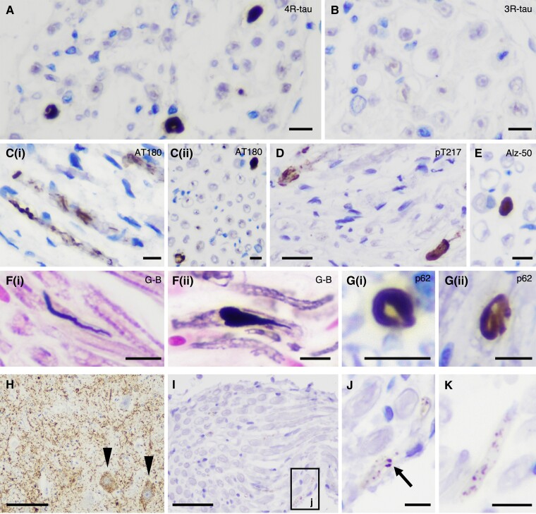
Figure 2.
Staining profiles of PNS-tau in progressive supranuclear palsy (PSP) and comparing morphological features of tau-positive inclusions in PSP and corticobasal degeneration (CBD). (A–G) Representative photomicrographs of staining profiles of the PNS-tau in PSP. The tau-positive inclusions in the PNS are detected with 4-repeat tau specific antibody (RD4) (A) but not with 3-repeat tau-specific antibody (RD3) (B). The tau-positive inclusions are also recognized with several phosphorylated tau antibodies [C(i and ii): AT180 and D: pThr217] other than AT8 and also Alz-50, which is for misfolded tau (E). The inclusions in PSP often show large structures with fibrillary morphology (A and E), which are stained with Gallyas–Braak silver staining (G-B) [F(i and ii)]. Some inclusions also show p62-immunoreactivity [G(i and ii)]. (H–K) Representative photomicrographs of tau pathology in the PNS and related nuclei in CBD Case 3, namely anterior horn (H), spinal anterior roots (I and J: high magnification image of black box ‘j’ in I) in the cervical cord and IX/X nerve (K). Although this CBD case shows massive tau pathology in the anterior horn of the cervical cord (H, arrowhead: pre-tangle-like inclusions), there were only a few fine granular tau deposits in the spinal anterior roots (I and J). (K) A small number of granular tau deposits is present in the IX/X nerve. (A) RD4, (B) RD3, (C) AT180, (D) pThr217, (E) Alz-50, (F) G-B, (G) p62 and (H–K) AT8. Scale bars in A–C(i and ii), E, F(i and ii), G(i and ii), J and K = 10 μm, D = 20 μm, H = 100 μm and I = 50 μm.

