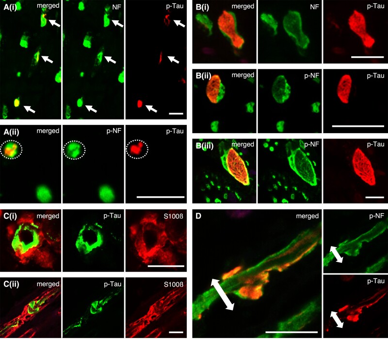
Figure 3.
Double-labelled immunofluorescence of phosphorylated-tau (p-tau) and neurofilament/S100β: the morphological features and localization sites of p-tau deposits in the peripheral nerve fibres. (A) In cross sections, phosphorylated tau deposits are primarily localized within the neuronal fibres (i)/axons (ii). (B) Some of the nerve fibres are filled with large tau inclusions showing fibrillary morphology (i–iii). (C) A part of the large tau-positive inclusions appear to accumulate in the lateral portion of nerve fibres, suggesting the possibility that the deposits are partly located within myelin. However, p-tau and S100β are not completely co-localized, and p-tau deposits merely appear to overlap on the myelin [C(i): cross and C(ii): sagittal section]. (D) Sagittal section of D-IF: p-tau and p-NF. In support of this, p-tau deposits with the outer portion are present within the axons (p-NF). In addition, the axons detected with p-NF seem to be bulged due to the large tau-positive inclusions. [A(i) and B(i)] NF (green) and pThr217 (red), [A(ii), B(ii and iii) and D] SMI31 (green) and pThr217 (red) and (C) AT8 (green) and S100β (red). Scale bars in A–D = 10 μm.

