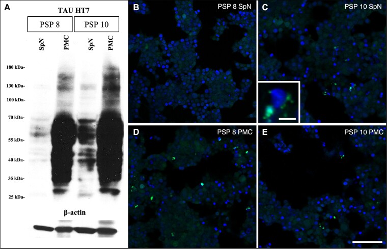
Figure 4.
Tau immunoblot analysis and in vitro tau seeding assay. (A) Tau immunoblot, total tau (HT7 clone), reveals the presence of tau in all four samples. (B–E) In vitro tau biosensor cells demonstrate that tau deposits (green) in the primary motor cortex (D and E) and cervical spinal nerve (C, enlarged image in the bottom left corner) samples have seeding capacity. (B and D) PSP Case 8 (PSP 8) and (C and E) PSP Case 10 (PSP 10). PMC = primary motor cortex; SpN = spinal nerves. Scale bars in B–E = 100 μm and C, enlarged image in the bottom left corner = 10 μm.

