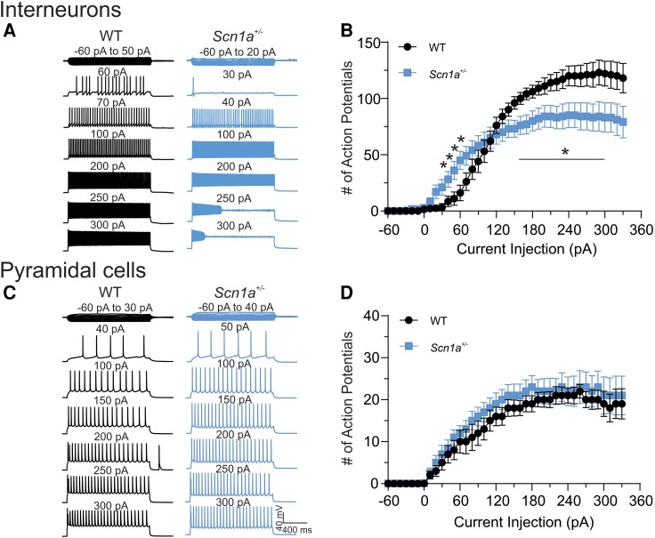
Figure 2.
Scn1a +/− PV+ interneurons, but not pyramidal neurons, show stimulation intensity-dependent biphasic changes in excitability. (A) Representative traces showing evoked repetitive firing of fast spiking PV+ interneurons in cortical layer 2/3 of brain slices from P21–25 wild-type (WT) (black) or Scn1a+/− (blue) mice. Repetitive action potential (AP) firing was evoked by injections of 1500 ms currents from −60 to 330 pA (selected responses are shown) at 10 pA-step from their resting membrane potentials. Here, a representative Scn1a+/− interneuron began to fire APs at lower intensities of current injection compared to the representative wild-type interneuron. Stronger depolarizing current injections blocked repetitive firing in the Scn1a+/− interneuron. (B) Input-output (I-O) curves for AP firing of wild-type versus Scn1a+/− PV+ interneurons in response to current injections. I-O curves were generated by plotting the number of evoked APs by 1500 ms current injections against current intensities over a range of −60 to 330 pA. (C) Representative traces showing evoked repetitive firing of pyramidal neurons in cortical layer 2/3 of brain slices from wild-type (black) or Scn1a+/− (blue) mice. Repetitive AP firing was evoked by injections of 1500 ms currents from −60 to 330 pA (selected responses are shown) at 10 pA-step from their resting membrane potentials. (D) I-O curves for AP firing of wild-type versus Scn1a+/− pyramidal neurons in response to current injections. I-O curves were generated by plotting the number of evoked APs by 1500 ms current injections against current intensities over a range of −60 to 330 pA. Values are mean ± standard error of the mean (SEM) of 25 cells from 15 wild-type mice or 26 cells from 13 Scn1a+/− mice, respectively. *Significant differences between wild-type and Scn1a+/− mice (P < 0.01). PV = parvalbumin.

