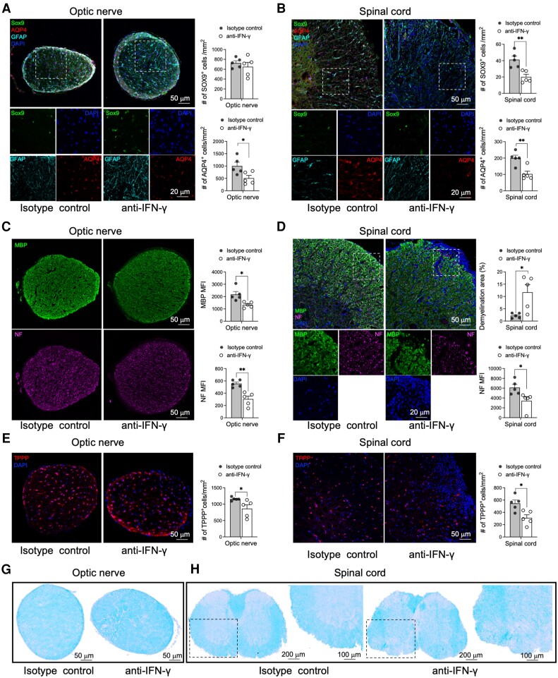
Figure 2.
AQP4201–220-induced disease is characterized by loss of astrocytes, oligodendrocytes, myelin and axons. Optic nerve (A, C and E) and lumbar spinal cord (B, D and F) sections were obtained from isotype control- and anti-IFN-γ-treated mice at Day 22 post-immunization (p.i.) and immunostained for astrocyte (Sox9-AQP4-GFAP-DAPI) (A and B), myelin (MBP-DAPI), neurofilament (NF-DAPI) (C and D) and oligodendrocyte (TPPP-DAPI) markers (E and F). The immunofluorescent images were accompanied by a statistical comparison of the markers’ mean fluorescence intensity (MFI) between the isotype control- and anti-IFN-γ-treated groups. Luxol fast blue (LFB) staining was performed to examine optic nerve (G) and lumbar spinal cord (H) for evidence of demyelination. The rectangle delineates an area of the anterior spinal columns that demonstrates loss of LFB staining depicted at low and high magnifications. Scale bars of 20, 50, 100 and 200 μm are indicated on the images. All data are presented as mean ± SEM (n = 5). Statistical analyses were performed using the Mann–Whitney test. *P < 0.05, **P < 0.01.

