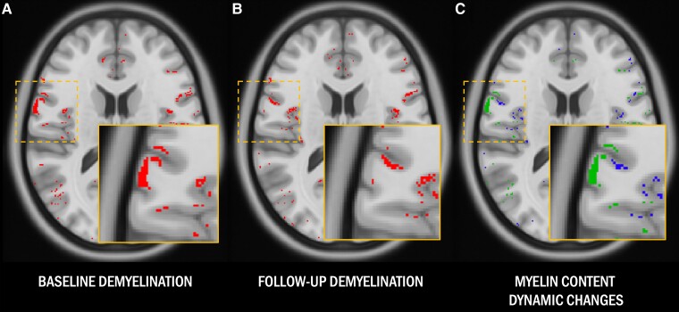
Figure 2.
Magnetization transfer ratio-based myelin content maps. (A and B) Cortical voxels in patients classified as demyelinated compared with healthy controls at baseline and follow-up are highlighted in red. (C) Cortical voxels classified as dynamically demyelinated (i.e. voxels classified as normally myelinated at baseline and demyelinated at follow-up) are highlighted in blue, while cortical voxels classified as remyelinated (i.e. voxels classified as demyelinated at baseline which recovered a normal magnetization transfer ratio signal at follow-up) are displayed in green.

