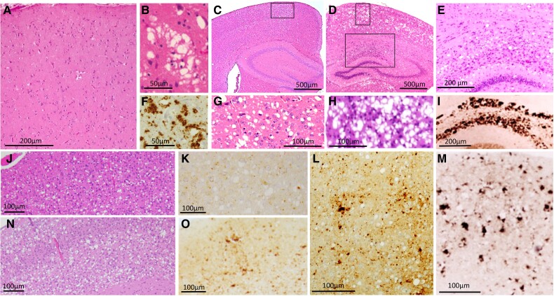
Figure 3.
Histopathology of the human sporadic fatal insomnia and of TgGlyc+ and TgGlyc− mice following inoculation of sporadic fatal insomnia isolates. Up to 18 months duration (A, B and F) typically there is minimal or no spongiform degeneration in the cerebral cortex (A), while beyond 2 year duration, spongiform degeneration and the PrP immunostaining pattern although focal mirror those of sCJDMM2 (B and F). In addition, sporadic fatal insomnia (sFI) typically presents with severe atrophy of anterior and dorsomedial thalamic nuclei similar to that of familial fatal insomnia (FFI) (see Fig. 4). (C, G and L) In TgGlyc+ mice (612 dpi), the spongiform degeneration was composed of mostly large and discrete vacuoles (C and G) and PrPD deposits of various sizes (L); (G: magnified region framed in C). TgGlyc− mice at first passage (260 dpi) (D, H and M) were affected by prominent and widespread spongiform degeneration and PrPD deposition that resembled, with a lower degree of severity, the histopathological features of the sCJDMM2-challenged TgGlyc− mice (D, H and M) (see also Fig. 1J). These similarities included the large PrPD aggregates of the hippocampus (E) forming the railway track-like PrP immunostaining pattern (I) (H, 90° left rotated and E are enlargements of the two regions framed in D). (J, K, N and O) The histotype of the two second passages with different incubation periods (77 and 227 dpi), showed spongiform degeneration with fewer and apparently smaller vacuoles, associated with lesser PrPD deposition in mice with the shorter incubation (J and K) compared with those where incubation was longer (N and O).

