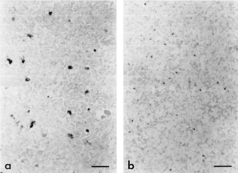
FIG. 2.
Photomicrograph of subadjacent sections from the spleen of a horse with acute clinical EIAVWSU5 infection, comparing the number cells containing viral RNA to the number of cells containing viral DNA. (a) Cells containing viral RNA (darkly staining cells) are labeled by ISH for EIAV gag RNA with the digoxigenin-labeled antisense probe and BCIP-NBT. Bar = 50 μm. (b) Cells containing viral DNA (darkly staining cells) are labeled by in situ PCR followed by ISH for EIAV gag cDNA with the digoxigenin-labeled sense probe and BCIP-NBT. Bar = 50 μm. Note the similar numbers of labeled cells in the sections, indicating that most or all infected cells are replicating virus during clinical disease.

