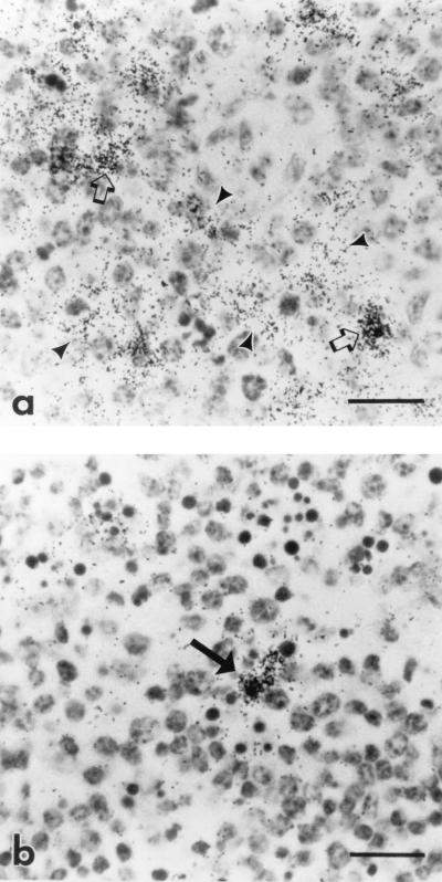
FIG. 3.
Photomicrograph of spleen and lymph node from a horse with acute clinical EIAV infection. Cells containing viral RNA (dark silver grains) are labeled by ISH for EIAV gag with the 35S-labeled antisense probe. (a) Germinal center in the spleen. Note the reticular pattern of silver grains indicating the presence of virus on the processes of follicular dendritic cells (arrowheads), in addition to the discrete pattern of silver grains indicating infection of individual cells (arrows). Bar = 20 μm. (b) Germinal center in the lymph node. Note the discrete pattern of silver grains indicating infection of individual cells (arrow) and absence of trapping of EIAV by dendritic cells. Bar = 20 μm.

