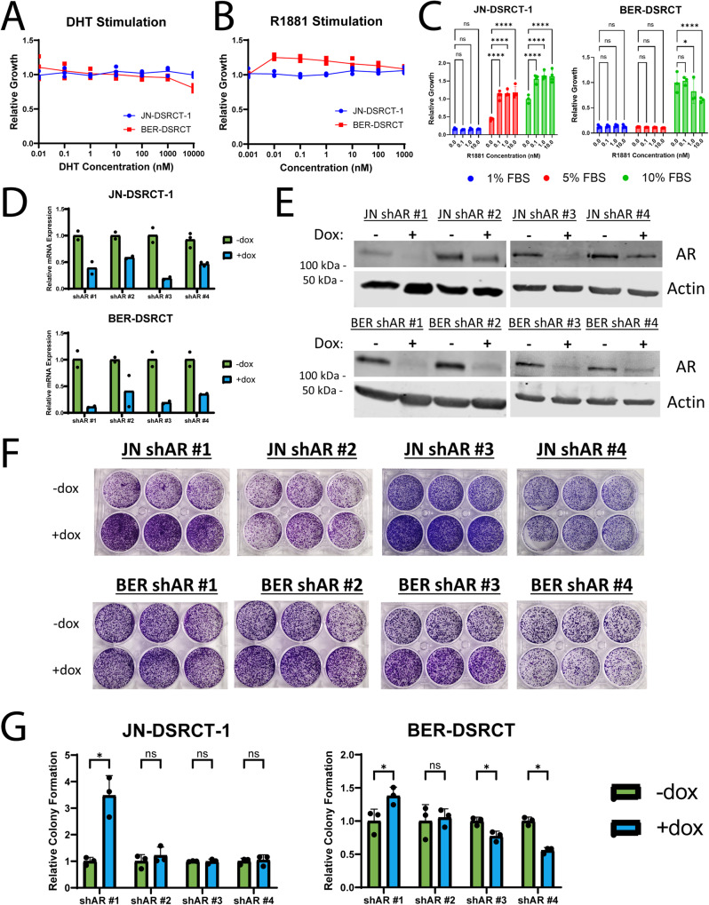
Fig. 5. DSRCT growth is independent of AR expression.
A Relative growth of JN-DSRCT-1 (blue) and BER-DSRCT (red) cells treated for 72 h with DHT (0.1 nM to 10 μM, n = 3 independent samples). B Relative growth of JN-DSRCT-1 (blue) and BER-DSRCT (red) cells treated for 72 h with R1881 (0.01 nM to 1 μM, n = 3 independent samples). C Relative growth of JN-DSRCT-1 and BER-DSRCT cells cultured in 1%, 5%, or 10% FBS and treated for 12 days with R1881 concentrations between 0.1 and 10 nM (n = 4 independent samples, *p < 0.05, ****p < 0.0001, Error bars = STD). D RT-qPCR of AR expression in JN-DSRCT-1 and BER-DSRCT shAR #1–4 cell lines with or without dox (n = 2–3 independent samples). E Western blot of AR protein expression in shAR #1–4 cell lines with or without dox (representative blot, n = 2 independent samples). F Images of colony formation assays from shAR #1–4 cell lines (n = 3 independent samples). G Quantification of colony formation assays of shAR #1–4 cell lines with (blue) or without dox (green) (n = 3 independent samples, *p < 0.05, **p < 0.01, Error bars = STD).

