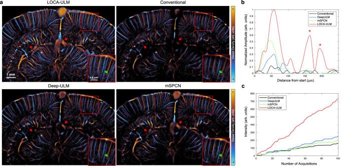
Fig. 5. Comparison among LOCA-ULM, conventional ULM, Deep-ULM14, and mSPCN21 in in vivo rat brain ultrasound data under high MB concentration (injection rate 40 μL/min).
a Each ULM image was generated by accumulating 25,000 frames of ultrasound data (a total of 100 s of acquisition, bregma: −5.6 mm) for MB injection rate of . b Intensity profile of vessels indicated by a white line in (a). c Accumulated intensity of reconstructed vessel with respect to an increased number of frames indicated by the white box in (a). n = 1 experiment. Source data are provided as a Source Data file.

