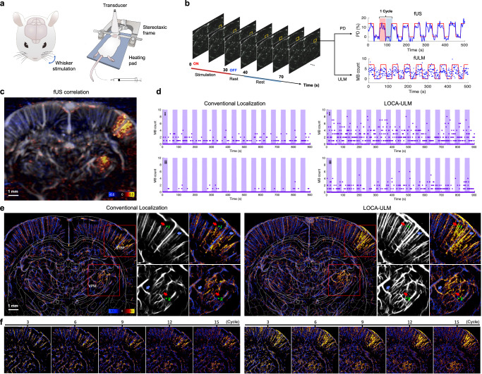
Fig. 6. LOCA-ULM increases MB signal sensitivity to blood flow during brain activation.
a, b Schematic of the experimental setup for functional ultrasound (fUS) and functional ULM (fULM) brain imaging conducted in the coronal plane (bregma: −3.3 mm). The fUS and fULM experiment involved whisker stimulation in an anesthetized rat with continuous intravenous injection of MBs (Methods). c fUS activation map calculated as the Pearson correlation coefficient between Power Doppler signals over time and the stimulation pattern. d MB count over time for a pixel in selected vessels (i and ii in e) comparing conventional ULM and LOCA-ULM. e fULM activation map calculated as the Pearson correlation coefficient between the MB count (after tracking) over time and the stimulation pattern for both conventional ULM and LOCA-ULM. Zoomed-in views of the ULM image and activation map in the barrel cortex (S1BF) and the ventral posteromedial nucleus (VPM) are shown, marked by a red rectangle. f Progressive enhancement of the fULM activation maps with an increasing number of stimulation cycle repetitions. n = 1 experiment. Source data are provided as a Source Data file.

