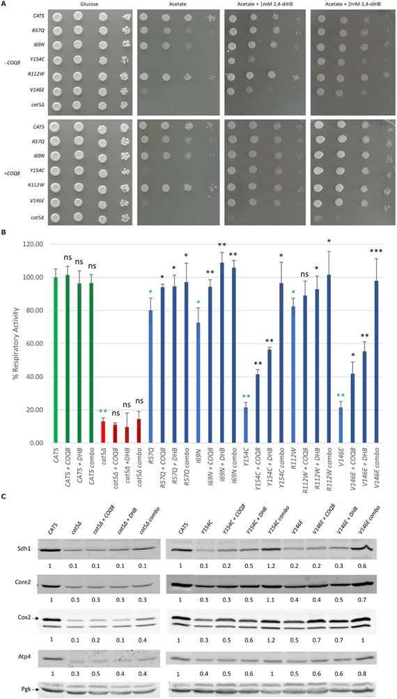
Figure 3. 2,4-Dihydrobenzoic acid supplementation and overexpression of COQ8 synergistically rescued oxidative growth defect of mutant cat5Δ strain.
(A) Oxidative growth: the cat5Δ strain harboring wild-type and mutant alleles or the empty vector with or without COQ8 overexpression (± COQ8) were serially diluted and spotted on SC agar plates supplemented with the fermentable carbon source 2% glucose or the non-fermentable carbon source 2% acetate, with or without the addition of 2,4-dihydroxybenzoic acid (2,4-diHB) and incubated at 28°C (B) Respiratory activity: Non-treated strains, strains with COQ8 overexpression (+ COQ8), with 2 mM 2,4-diHB supplementation (+ DHB) or with both treatments (combo) were grown at 28°C in SC medium without uracil and leucine supplemented with 0.6% glucose. Values are represented as the mean of at least three values ±SD. The green bars indicate the wild-type strains; the blue bars indicate strains carrying different COQ7 variants; the red bars indicate the null mutant strains. Light colors for the non-treated strains, and dark colors for the treated strains. Statistical analysis was performed using a one-tail paired Student’s t-test comparing the non-treated mutant strains with the non-treated wild type strain (green asterisks) or comparing the treated strains to the relative non-treated strain (black asterisks/ns). ns: not significant, * p<0.05, ** p<0.01, *** p<0.001. (C) Representative Western blots for the evaluation of respiratory complexes subunits levels on the wild-type strain, cat5Δ strain and strains carrying Y154C and V146E variants with COQ8 overexpression (+ COQ8), 2,4-diHB supplementation (+ DHB) and with both the treatments (combo). Antibodies used: Sdh1 for complex II; Core2 for complex III; Cox2 for complex IV; Atp4 for complex V; Pgk as loading control. The quantification was performed on two independent blots using Image Lab Software (Bio-Rad).

