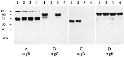
FIG. 2.
Protein profile of mutant PrV. Virions of wild-type PrV (lanes 1), PrV-gC− (lanes 2), PrV gD− Pass (lanes 3), and PrV gCD− Pass (lanes 4) were lysed, and proteins were separated by acrylamide gel electrophoresis. After transfer to nitrocellulose membranes, the filters were probed with MAbs specific for gB, gC, gD, and gH (α gB, αgC, αgD, and αgH, respectively). Bound antibody was visualized after incubation with peroxidase-conjugated secondary antibody by enhanced chemiluminescence recorded on X-ray film. The anti-gB antibody recognizes the uncleaved precursor as well as one of the two proteolytic cleavage products.

