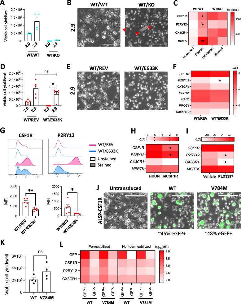
Fig. 7.
Effect of CSF1R loss of function on microglial phenotype. A-C CSF1RWT/WT and CSF1RWT/KO iPSCs were differentiated into iMGL side-by-side. A Viable cell yield per well of a 6-well plate assessed by trypan blue exclusion assay following 2.0 and 2.9 differentiation. A Kruskal–Wallis test was performed, followed by Dunn’s post hoc test. n = 4 differentiation batches. B Phase contrast images of iMGL 2.9. Red triangles show presence of round or dysmorphic floating cells. Scale bar = 150 . C Flow cytometry assessment of cell surface marker expression. Kruskal–Wallis tests were performed, followed by Dunn’s post hoc test. n = 4 differentiation batches using the 2.9 protocol, * p < 0.05, ** p < 0.01 vs. unstained control. MFI = median fluorescence intensity, a.u. = arbitrary unit. D-G CSF1RWT/REV and CSF1RWT/E633K iPSCs were differentiated into iMGL side-by-side. D Viable cell yield per well of a 6-well plate assessed by trypan blue exclusion assay following 2.0 and 2.9 differentiation. A Kruskal–Wallis test was performed, followed by Dunn’s post hoc test. n = 4 differentiation batches. E Phase contrast images of iMGL 2.9. Scale bar = 150 . F qRT-PCR assessment of microglia markers. T-tests were performed. n = 5 differentiation batches using the 2.9 protocol, * p < 0.05 vs CSF1RWT/REV iMGL. G Flow cytometry assessment of cell surface marker expression. Mann–Whitney tests were performed. n = 5 differentiation batches using the 2.9 protocol * p < 0.05, ** p < 0.01. MFI = median fluorescence intensity. H-I Healthy control, mature iMGL were generated following the 2.9 protocol. H Microglia marker expression assessed by qRT-PCR in iMGL three days after siCON or siCSF1R transfection. T-tests were performed. n = 3 lines, * p < 0.05 vs siCON. I Microglia marker expression assessed by qRT-PCR in iMGL following a 3-day treatment with PLX3397 (1 μM, every other day). T-tests were performed. n = 4 lines, * p < 0.05 vs vehicle treatment. J-L eGFP and either WT or V784M CSF1R were stably co-expressed in ALSP-CSF1R iHPCs using lentiviruses and cells were differentiated into iMGL following the 2.9 protocol. J Merged phase contrast and green fluorescence images of ALSP-CSF1R iMGL. Scale bar = 150 μm. (K) Viable cell yield per well of a 6-well plate assessed by trypan blue exclusion assay. A t-test was performed. n = 4 differentiation batches. L Flow cytometry assessment of eGFP signal and microglia marker expression. MFI = median fluorescence intensity. n = 1 differentiation batch

