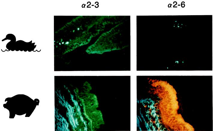
FIG. 1.
Comparison of lectin staining in duck intestine (colon) and pig trachea. The M. amurensis lectin specific for NeuAcα2,3Gal (designated α2,3; detected with fluorescein isothiocyanate-labeled anti-DIG antibody) bound to both duck intestinal epithelium and pig tracheal epithelium, whereas S. nigra lectin specific for NeuAcα2,6Gal (designated α2,6; detected with rhodamine-labeled anti-DIG antibody) bound only to the latter. Blue staining in the connective tissue represents autofluorescence. Magnification, ×300.

