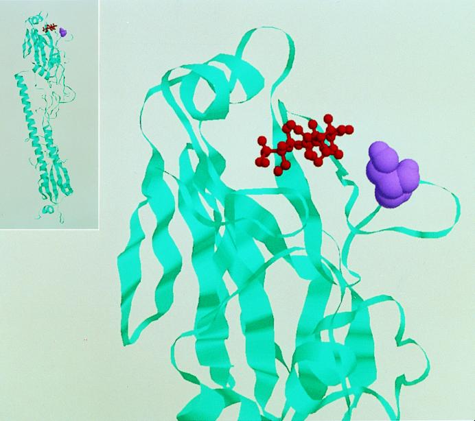
FIG. 4.
Globular head of the influenza virus HA molecule (ribbon, cyan), illustrating the location of Ser145 (space-filled, purple) relative to that of bound sialic acid (ball and stick, red). Inset shows the entire molecule. This figure is based on the H3 HA structure of A/Aichi/2/68 (H3N2) complexed with sialic acid as determined by Weis et al. (40) and is not intended to represent the actual three-dimensional structure of the H1 molecule.

