Figure 3.

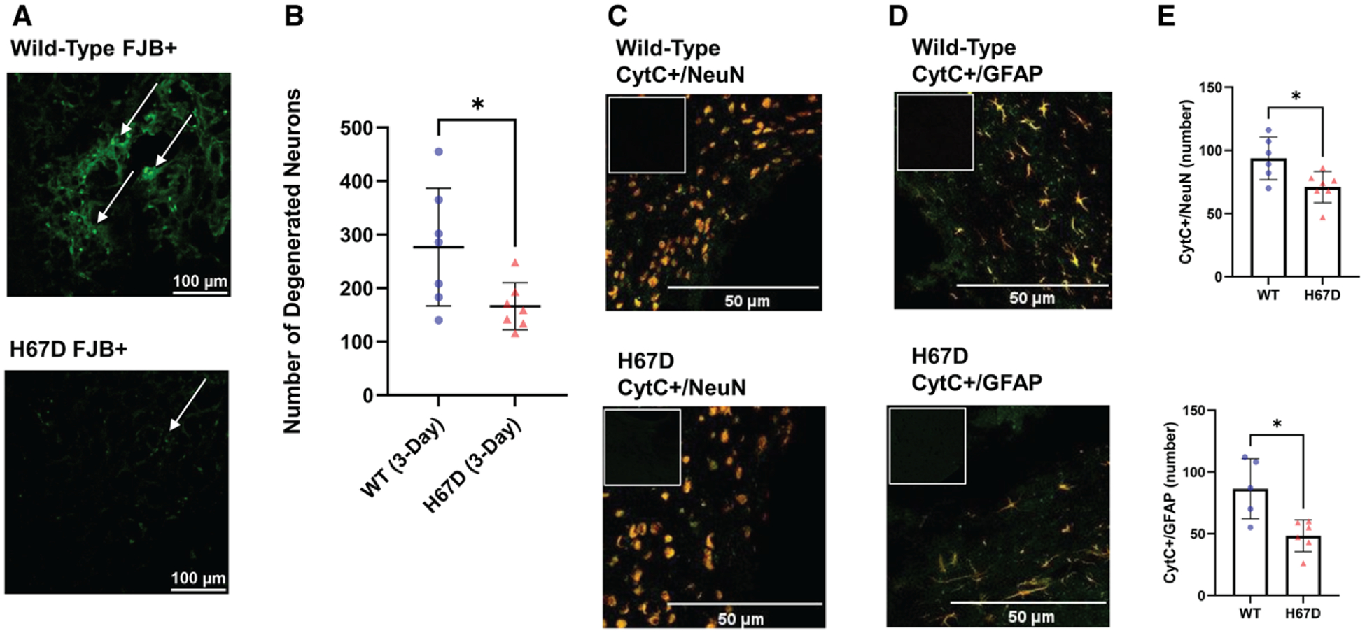
The neuroprotective effects of the H67D mice following intracerebral hemorrhage (ICH).
Fluorojade-B (FJB) staining determined differences in the number of degenerated neurons in the perihematomal region in (A) wild-type (WT) mice (top) and H67D mice (bottom) at 3 d following ICH; arrows point to FJB-positive cells. B, Quantification of FJB-positive cells done using ImageJ. The H67D showed significantly decreased FJB-positive cells (n=7, mean, 166.1, SD=44.02) in the perihematomal region at 3 d post-ICH compared with WT mice (n=7, mean, 277, SD=109.9; P=0.0291). CytC (cytochrome C; green) and NeuN ([neuronal nuclear protein]; red) colocalization in the perihematomal area of (C) WT mice (top) and H67D mice (bottom) at 3 d following ICH; CytC (green) and GFAP ([glial fibrillary acidic protein]; red) colocalization in the perihematomal area in (D) WT mice (top) and H67D mice (bottom); (E) Quantification of CytC+ NeuN cells and CytC+ GFAP cells. H67D showed significantly fewer CytC+ NeuN cells (n=7, mean, 71, SD=12.26) in the perihematomal region at 3 d post-ICH compared with WT mice (n=6, mean, 93.67, SD=16.81; P=0.0210). H67D also showed significantly fewer CytC+ GFAP cells (n=6, mean, 48.33, SD=12.83) in the perihematomal region at 3 d post-ICH compared with WT mice (n=5, mean, 85.40, SD=24.38; P=0.0216). Respective negative immunofluorescent staining is displayed in upper left corner. Error bars reported as SD, *P<0.05.
