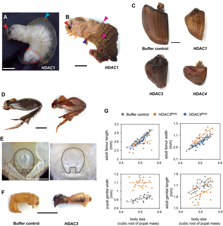
Fig. 2.
HDACRNAi effects on molting and appendage development. A and B Larval–pupal intermediate induced by HDAC1 knockdown. The fat body accumulated outside of newly formed pupa is outlined (red dotted line) before (A) and after (B) peeling away the larval cuticle, respectively. Hemolymph in the cavity between larval and newly formed pupal cuticle, pupal compound eyes, wings, and pupal support structures are indicated by cyan, red, blue, and magenta arrowheads, respectively. C Representative wing phenotypes are shown as follows: buffer injection, HDAC1RNAi, HDAC3RNAi, and HDAC4RNAi. D-F Morphology of the hind leg (D), pupal genitalia (E), and adult aedeagus (F) compared to buffer-injected (left column) and HDAC3RNAi (right column) individuals, respectively. G Knockdown of HDAC3 (orange dots) or HDAC4 (blue dots) on femur length and width compared to buffer-injected control (gray dots). Effects of HDAC3RNAi on the male pupal and adult reproductive organ (bottom row in G). The RNAi phenotypes and their corresponding negative controls are shown at the same magnification. Scale bars: 1 mm

