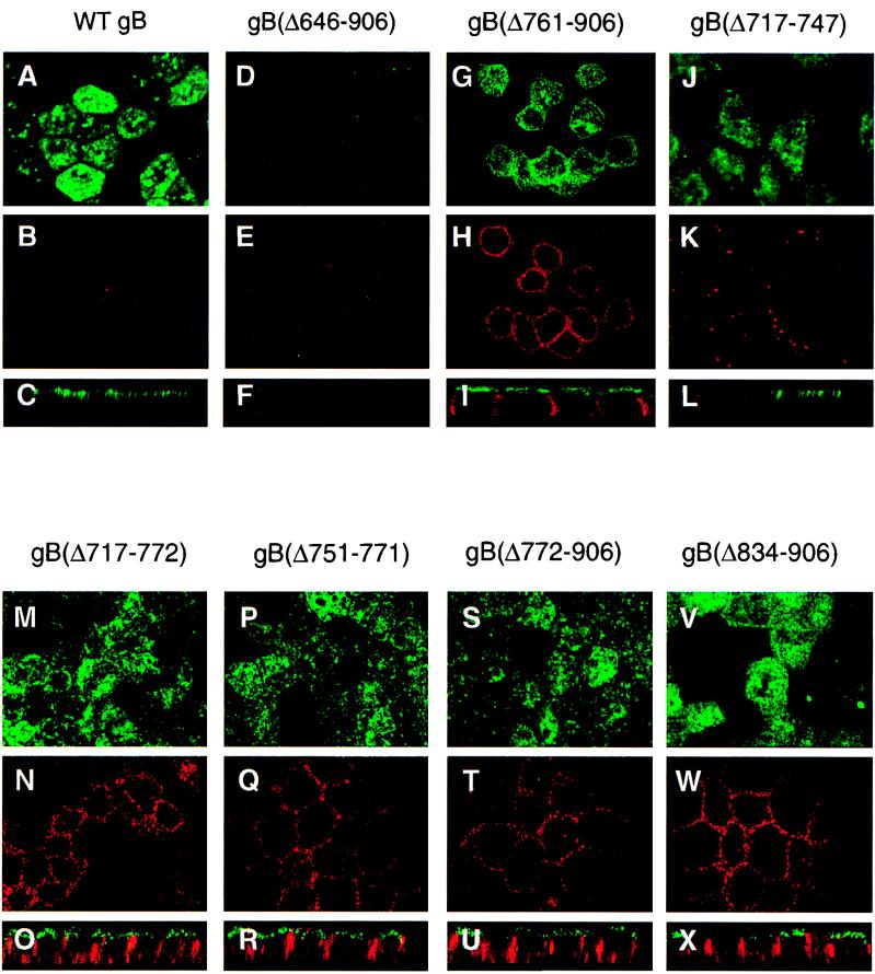
FIG. 3.
Immunofluorescence confocal microscopy showing the transport of CMV WT gB and deletion derivatives to the apical and basolateral surface membranes of polarized MDCK cells. Cells were incubated with a pool of MAbs to gB, fixed, and then reacted with secondary antibodies conjugated either with fluorescein isothiocyanate (AP membrane) or with Texas red (BL membrane). Derivatives are indicated at the top of each set of panels.

