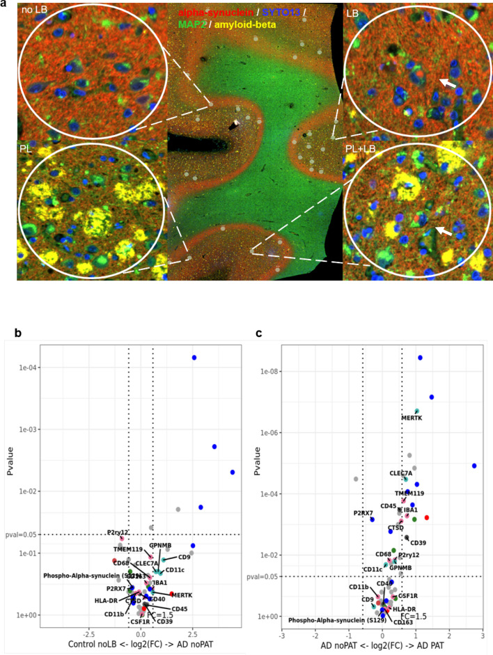
Fig. 8.
Analysis of microenvironment around neurons with Aβ plaque pathology in cases with ADNC (AD and MIX) and Controls in frontal cortex. a Selected region of interests (ROIs) from tissue with ADNC (AD, MIX) and Control. For the selection of pathology, circles of 200 μm were drawn around neurons with and without Aβ plaque. Morphology markers α-Syn (NanoString α-Syn Alexa Fluor® 594) (red), α-MAP-2 (NanoString Alexa Flour® 532) (green) and beta-amyloid (D54D2) (Alexa (R) 647) (yellow) were used to distinguish neurons with accumulations of α-Syn in form of Lewy bodies (arrows) and Aβ plaques. DNA was stained with SYTO13 dye (blue). b Volcano plot for the expressed proteins around neurons without pathology in cases with Aβ pathology (AD, MIX) and Controls. c Volcano plot for the expressed proteins around neurons with and without pathology in disease cases (AD, MIX), comparison of the microenvironment of pathology and non-bearing neurons within the same tissue. In volcano plots, unadjusted p‐value of 0.05 and fold‐change (FC) of 1.5 were used to identify differentially expressed proteins. Color code of detected proteins based on used NanoString Antibody Panels: blue – AD, red – Astrocyte/Inflammation, green – PD, pink – Microglia, turquoise – Disease-Associated Microglia, dark grey – Microglia/Inflammation, light grey – other

