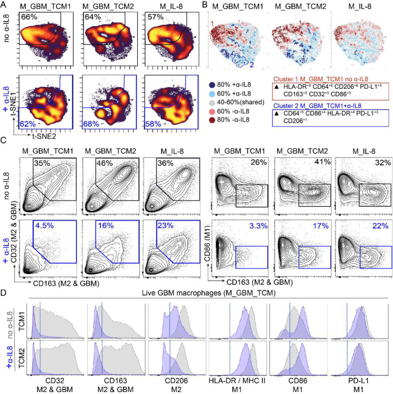
Figure 3 – GBM secreted IL-8 mediates ex vivo polarization of macrophages to express a suppressive signature.
A) t-SNE plots display cell density of polarized macrophages across different conditions including recombinant IL-8 (M_IL-8) and GBM tumor conditioned media (M_GBM_TCM), in the presence or absence of α-IL-8 blocking antibody. B) T-REX analysis comparing polarization conditions in the presence or absence of α-IL-8 where cells that are >80% enriched in macrophages polarized in the absence of α-IL-8 are colored in dark red, >60% enriched in light red, cells enriched >80% in macrophages polarized in the presence of α-IL-8 are colored in dark blue and >60% enriched in light blue. Cells colored in gray are similarly enriched in both conditions C) 2D contour plots show expression of signature surface proteins CD163, CD32, CD86, and PD-L1 for each condition. Gates colored in blue indicate the conditions where macrophages were polarized in the presence of α-IL-8. D) Histogram plots display individual protein expression of markers CD32, CD163, CD206, HLA-DR, CD86 and PD-L1. Overlaid histograms represent macrophages polarized in the presence (blue) or absence (gray) of α-IL-8 blocking antibody. A blue dotted line indicates a threshold for positive expression.

