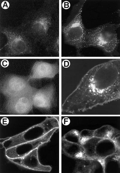
FIG. 7.
Transfection of Us9 constructs. PK15 cells were grown on glass coverslips and transfected by the calcium phosphate method with pAB7 (A), pBB14 (B, D, E, and F), or EGFP (C). At 72 h posttransfection, Us9 was detected by indirect immunofluorescence microscopy with Us9 antiserum (A) as described in the legend to Fig. 5. The localization of pBB14 (B, D, E, and F) and EGFP (C) was determined at 48 h posttransfection by confocal microscopy under UV illumination. Panel D is a higher magnification of the Us9-EGFP-transfected cells shown in panel B, clearly demonstrating expression of the fusion protein in the Golgi compartment and plasma membrane. (E and F) Sensitivity of Us9-EGFP (pBB14) to BFA treatment. PK15 cells transfected with pBB14 were treated with 2.5 μg of BFA per ml for 60 minutes (E). Some BFA-treated cells were then washed three times with fresh medium and allowed to recover for 1 h (F).

