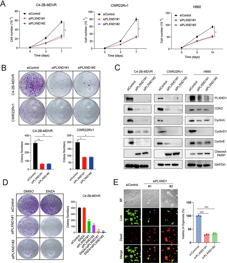
Figure 3. Knockdown of PLXND1 represses the cell proliferation and improves enzalutamide treatment.
A. C4–2B-MDVR (1*10^4 cells/well), CWR22Rv1 (1*10^4 cells/well), and H660 (5*10^4 cells/well) cells were plated in 12-well plates and transfected with siControl or siPLXND1 siRNAs, respectively. Cell proliferation viability was determined by cell counting. B. C4–2B-MDVR and CWR22Rv1 cells were plated in 6-well plates, 1000 cells/well, and colony formation viability was determined by counting after transfected with siControl or siPLXND1 siRNAs for 10 days. C.C4–2B-MDVR, CWR22Rv1, and H660 cells after transfected with siControl or siPLXND1 siRNAs for 5 days. Then, whole cell lysates of C4–2B-MDVR, CWR22Rv1, and H660 cells were harvested for testing PLXND1 and other proteins expression levels. D. C4–2B-MDVR cells were plated in 6-well plates, 1000 cells/well, and colony formation was determined by counting after transfected with siControl or siPLXND1 siRNAs with or without 20 μM enzalutamide for 10 days. E. Cells from H660 PDX tumors were plated in 96-well plates, 1*10^4 cells/well, and transfected with siControl or siPLXND1 siRNAs for 10 days. Organoids viability was assayed by CellTiter-Glo Luminescent assay and the live-and-dead cells were visualized by immunofluorescence. * p<0.05, **P<0.01, ***P < 0.001.

