FIG. 6.
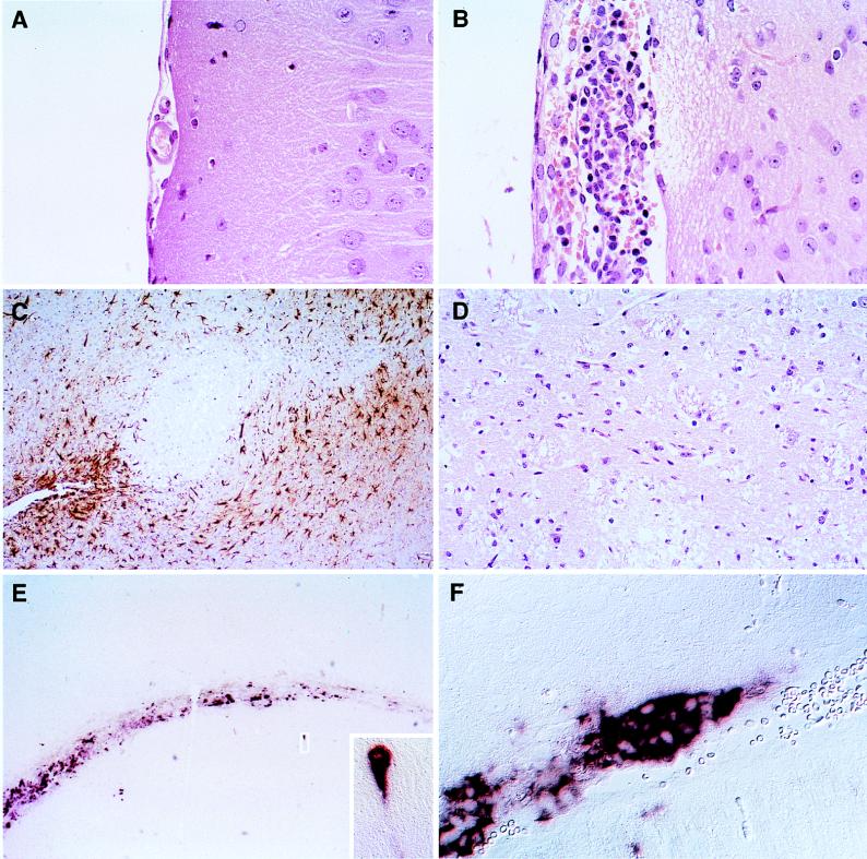
Histological analysis of the consequences of MV infection and of the MV RNA distribution in the brains of Ifnartm-CD46Ge animals. A mouse inoculated with 1 million infectious units of MV-Edm (B to F) and a mock-infected mouse (A) were sacrificed 3 days p.i., their brains were prepared, and brain slices were stained as indicated below. (A, B, and D) HE-stained sections showing meningeal inflammation (B) and necrosis (B and D) of the brain tissue. (C) Immunohistochemical staining for GFAP demonstrating reactive astrocytes (brown) surrounding necrotic lesions. (E and F) In situ hybridization with a MV N-specific probe showing strong staining in the ventricular region and in scattered neurons. One infected hippocampal neuron is enlarged in the inset. In panel F, many contiguous ependymal cells are stained.

