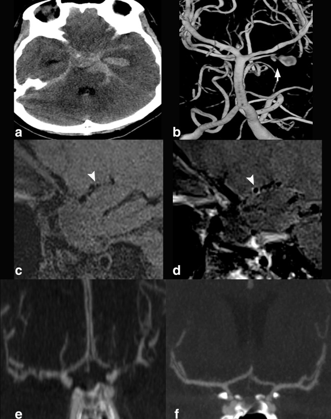
Figure 5.
25 year old female who presented with severe headache and altered mental status. On axial non-contrast CT head (A), there was diffuse basilar subarachnoid hemorrhage and intraventricular blood in the left temporal horn. 3D rotational angiogram of the left vertebral artery (B), demonstrated posteriorly directed, lobulated left superior cerebellar artery aneurysm (short arrow). After the ruptured aneurysm was treated with coil embolization, sagittal T 1-weighted pre- (C) and post-contrast (D) HR-VWI demonstrated right M1 MCA segment wall enhancement. This finding is significantly associated with subsequent angiographic vasospasm. CTA coronal MIP (E) obtained two days after HR-VWI performance, when compared to initial presentation CTA coronal MIP (F), showed diffuse angiographic vasospasm.

