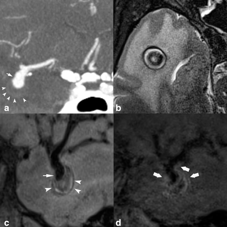
Figure 6.
48 year old with partially thrombosed right MCA aneurysm. Coronal MIP CTA reconstruction (A) shows patent aneurysm lumen projecting inferiorly from the MCA trifurcation (short arrow), with subtle boundary representing the margin of the thrombosed aneurysm sac (arrowheads). On axial T 2-weighted HR-VWI (B), aneurysm sac thrombus shows heterogeneous signal with central T2 hypointensity and peripheral hyperintensity. On sagittal T 1-weighted HR-VWI (C), patent aneurysm lumen projecting inferiorly from the MCA (short arrow), with inferior aneurysm heterogeneous intraluminal thrombus (arrowheads) are appreciated. On sagittal post-contrast T 1-weighted HR-VWI (D), there is AWE (thick arrows).

