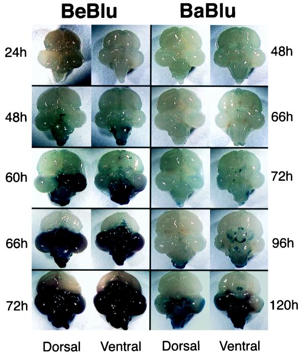
FIG. 5.
Progression of PRV infection in the chicken embryo brain. At the indicated times after injection into the right eye, the brains from infected embryos were removed and placed in X-Gal. Only infected tissue close to the surface of the brain or exposed by necrosis was stained blue. Dorsal and ventral views of each brain are shown. Note that the times that the brains were collected are different for Be-Blu- and Ba-Blu-infected embryos. It is important to note that many of the BeBlu-infected animals sacrificed at 48 h had more extensive infection than the examples illustrated and that in some cases their brains resembled the 66-h brain. This inherent variability is likely due to the use of genetically outbred animals but nevertheless emphasizes the speed with which BeBlu infection exerts its pathology.

