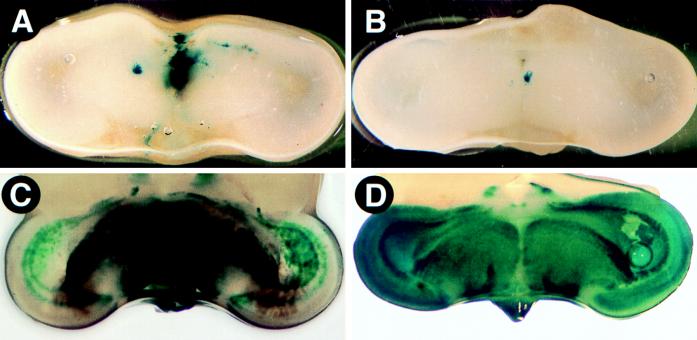
FIG. 7.
PRV infection of the midbrain after intraocular inoculation. The brains of infected embryos were sliced in the coronal plane through the midbrain by using a razor blade and placed in X-Gal substrate buffer for 1 h. (A) Section through the midbrain of a BeBlu-infected embryo at 48 h postinfection; (B) section through the midbrain of a BaBlu-infected embryo at 48 h postinfection; (C) section through the midbrain of a BeBlu-infected embryo at 60 h postinfection; (D) section through the midbrain of a BaBlu-infected embryo at 96 h postinfection.

