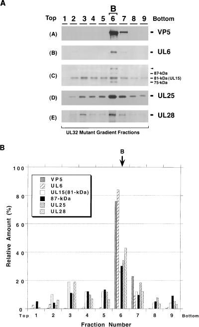
FIG. 10.
Sucrose gradient analysis of lysates from cells infected with hr64 lacking UL32. Gradients similar to those described in the legend to Fig. 3 were collected into nine fractions. Fraction 6 contains B capsids. (A) Immunoblot analysis was performed to detect VP5 (panel A), UL6 (panel B), UL15 (panel C), UL25 (panel D), and UL28 (panel E). A long (20 min) exposure of the UL15 immunoblot is shown. (B) Quantitative analysis was performed as described in the legend to Fig. 3.

