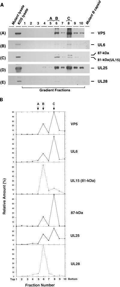
FIG. 3.
Sucrose gradient analysis of lysates from KOS-infected cells. KOS-infected cell lysates were fractionated by ultracentrifugation on a 15 to 45% (wt/wt) sucrose gradient (in phosphate buffer) as described in Materials and Methods. Ten fractions from the gradient were collected and examined for individual proteins as indicated. (A) Gradient fractions were examined by immunoblotting with the appropriate antibodies for VP5, UL6, UL15, UL25, and UL28 as indicated. The UL15-related bands were revealed by exposure for 20 min. KOS lysate represents a total-cell lysate from KOS-infected cells; Mutant lysate represents a mock-infected-cell lysate (A), or a total-cell lysate from cells infected with the mutant virus defective for UL6 (B), UL15 (C), UL25 (D), or UL28 (E); Mutant B capsid represents the B capsids isolated from cells infected with a mutant virus defective for UL6 (B), UL15 (C), UL25 (D), or UL28 (E). (B) Quantification of the immunoblots from Fig. 3A was carried out as described in Materials and Methods. The y axis shows the relative amount of each protein in each fraction calculated as a percentage of the total amount of protein. The total amount of protein was calculated by summing the amount of protein in each fraction. Fractions 5, 6, and 8 contain the A, B, and C capsids, respectively. The top and bottom of the gradient are indicated.

