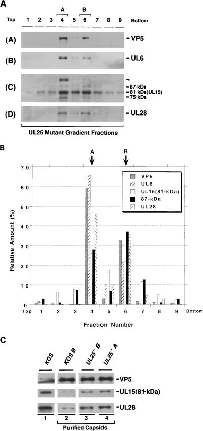
FIG. 8.
Sucrose gradient analysis of lysates from cells infected with KUL25NS lacking UL25 (UL25−). Gradients similar to those described in the legend to Fig. 3 were collected into nine fractions. Fractions 4 and 6 contain A and B capsids, respectively. (A) Immunoblot analysis was performed to detect VP5 (panel A), UL6 (panel B), UL15 (panel C), and UL28 (panel D). A long (20 min) exposure of the UL15 immunoblot is shown. (B) Quantitative analysis was performed as described in the legend to Fig. 3. (C) Analysis of the 81-kDa UL15 and UL28 levels in capsids from cells infected with the wild type or KUL25NS. A or B capsids were purified by sucrose gradient sedimentation. The amount of capsids in each sample was determined by normalization to the amount of VP5 present. Roughly equal amounts of capsids were subjected to immunoblot analysis with antisera against VP5, UL15, and UL28. Lysates of cells infected with KOS were also included to indicate the position of each of the proteins (lane 1).

