. 2024 Apr 5;12:RP89303. doi: 10.7554/eLife.89303

Figure 7. PARG is essential for cell survival.

(a) Diagram of full-length PARG was presented with the indicated gRNAs (gRNA#3 and 4) which target different regions in the C-terminal catalytic domain. The boundary of the catalytic domain was depicted based on Uniprot annotation. gRNA#1 and gRNA#2 were used previously to generate the aforementioned HEK293A- and HeLa-derived PARG KO cells, respectively; while gRNA#3 and gRNA#4 were used to generate PARG complete/conditional knockout (cKO) in the presence of olaparib in HEK293A and HeLa cells. (b) Immunoblotting was conducted to confirm the loss of PARG in PARG cKO cells derived from HEK293A and HeLa cells, which were cultured in the presence of 100 nM olaparib. (c) Clonogenic assay results of WT and PARG cKO cells treated with or without PARPi (100 nM) for 7 days. (d) Left: The immunoblots to confirm reconstitution with WT PARG or catalytic inactivation PARG in HEK293A PARG cKO cells. Right: Results of clonogenic survival assay with HEK293A PARG cKO cells reconstituted with WT or catalytic inactivation mutant of PARG for 7 days. (e) Representative clonogenic results conducted in HEK293A PARG cKO cells treated with NAM (100 µM) or NMN (1 mM) for 7 days. (f) HEK293A PARG cKO cells were synchronized with double thymidine block (DTB). Cells remained with DTB or were released from DTB for 4 hr, and then fixed and stained with anti-pADPr antibody and FxCycle Violet dye. (g) Immunoblots of soluble and chromatin-bound PARP1 and pADPr levels in HEK293A WT and PARG cKO cells treated with DMSO or olaparib (10 µM) for 2 hr.

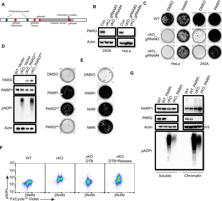
Figure 7—source data 1. Original file for the western blot analysis and colony formation assay in Figure 7.
Figure 7—source data 2. PDF containing Figure 7 and original scans of the relevant western blot analysis and colony formation assay with highlighted bands and sample labels.

Figure 7.

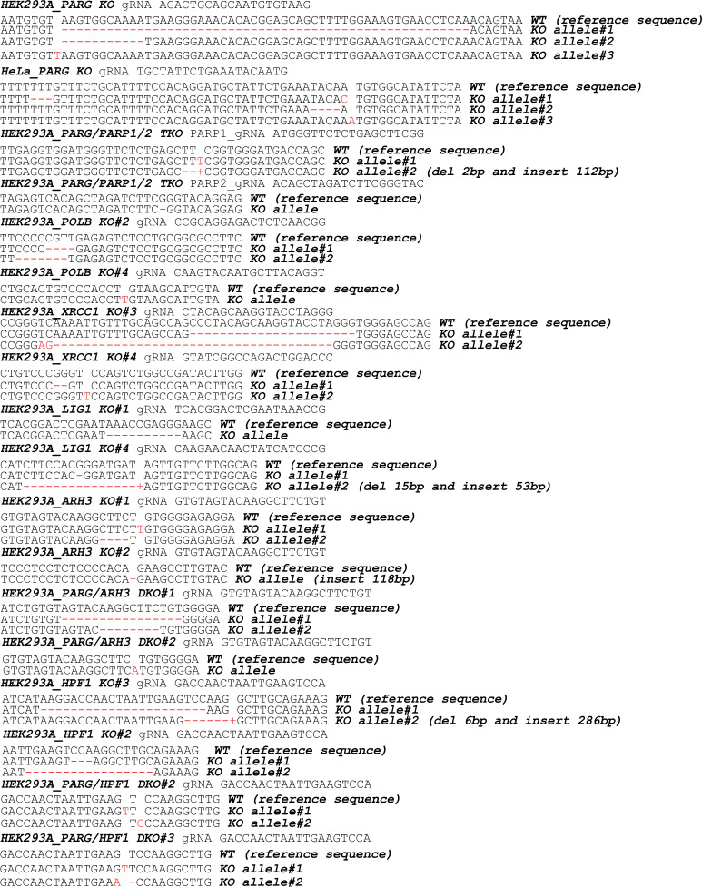
Figure 7—figure supplement 1. KO cells were validated by DNA sequencing.

Figure 7—figure supplement 1.

gDNA sequences were provided for each KO clones. The WT reference sequence was also provided.
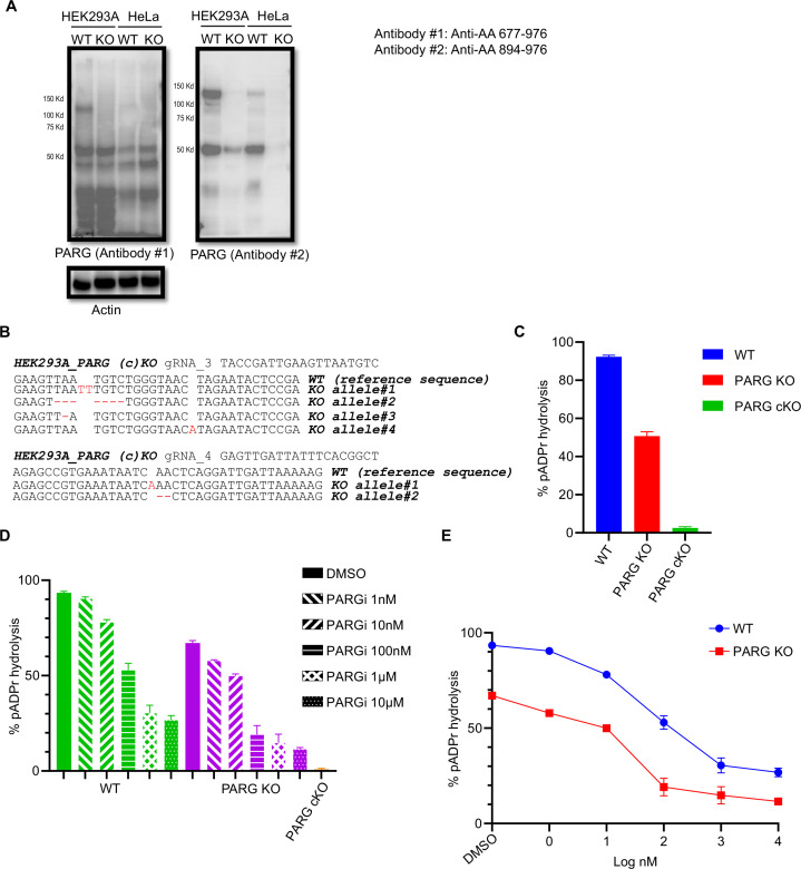
Figure 7—figure supplement 2. DePARylation activity of PARG is essential for cell survival.

Figure 7—figure supplement 2.

(a) Immunoblotting of PARG using two independent antibodies that recognize the C-terminus of PARG in PARG KO cells. The immunogens and the corresponding amino acid regions were shown (left). (b) HEK293A cKO cells were validated by DNA sequencing. gDNA sequences are indicated at the top. The WT reference sequence was provided. (c) The dePARylation activity was assessed in whole cell lysates (WCLs) prepared from control wild-type (WT), PARG KO, and PARG cKO HEK293A cells. The dePARylation activity was measured with remnant pADPr recognized by anti-pADPr antibody, and normalized with the Control and the Blank samples as indicated in Materials and methods. n=4 independent measurements. (d) The PARG dePARylation activity in WCL prepared from control WT and PARG KO HEK293A was inhibited in a dose-dependent manner in the presence of PARGi. WCL prepared from PARG cKO cells was included as a control. n=3 independent measurements. (e) The PARG dePARylation activity shown in (d) is presented together with PARGi concentrations.
Figure 7—figure supplement 2—source data 1. Original file for the western blot in Figure 7—figure supplement 2A.
Figure 7—figure supplement 2—source data 2. PDF containing Figure 7—figure supplement 2A and original scans of the relevant western blot analysis with highlighted bands and sample labels.