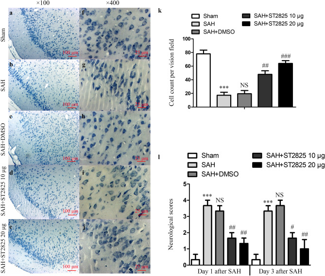
Figure 2.
ST2825 treatment ameliorated brain tissue damage and clinical neurological after SAH. (a–j) Representative slides of Nissl staining at two different magnifications (a–e, × 100, f–j, × 400) to visualize the neuronal cell outline and structure. SAH reduced the number of the neurons, and treatment of ST2825 preserved neurons from damage, including neuron loss and degeneration. Cells in the SAH and DMSO treated groups were arranged sparsely and the cell outline was fuzzy compared to sham group. (k) Cell counts per visual field (× 400) was quantified in the slides with Nissl staining. (l) Neurological assessment of SAH animals treated with DMSO or ST2825. In comparison with the control group, SAH significantly increased the neurological scores both at 24 h and 72 h post-SAH. ST2825-treated rats exhibited significant improvement in clinical behavioral function at both 24 and 72 h after injury when compared to the SAH rats or DMSO treated rats. Data are expressed as mean ± SD (n = 6 in each group). ***p < 0.001 compared with the sham group, #p < 0.05 compared with the SAH group, ##p < 0.01 compared with the SAH group, ###p < 0.001 compared with the SAH group, NS, no statistic difference compared with the SAH group.

