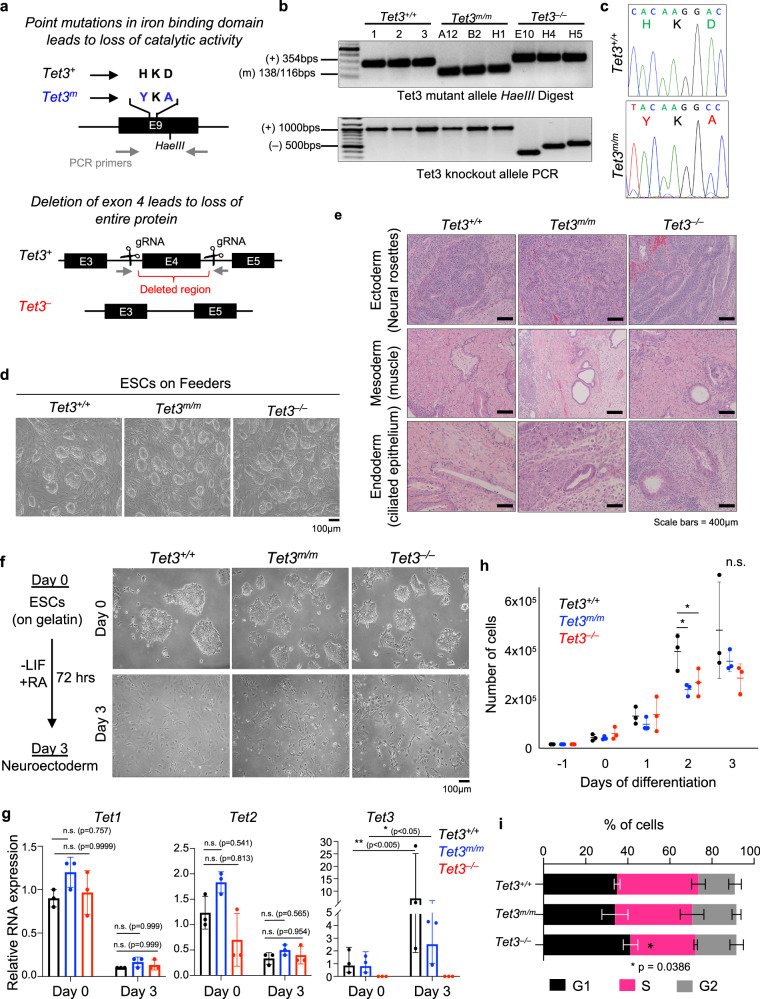
Fig. 1. Generation of Tet3m/m and Tet3–/– mouse ESC and their differentiation to NE.
a Schematic of the gene targeting strategy to generate catalytic mutant (Tet3m) and knockout (Tet3–) mouse ESCs. b Genotyping of Tet3m/m clones by RFLP (restriction fragment length polymorphism) using HaeIII enzyme (top). Correctly targeted (mutated) allele bands are 138 bp and 116 bp. Allele without mutation is 354 bp. (3 independent clones were generated A12, B2, and H1). Genotyping of Tet3–/– clones by PCR (bottom). Amplification of a shorter fragment (~500 bps) confirms deletion of exon 4 (3 independent clones were generated E10, H4, and H5). c Sanger sequencing confirming mutations for amino acid substitutions HKD to YKA in Tet3m/m ESCs. d Representative brightfield images of ESCs cultured on feeder cells. Scale bar = 100μm. e Hematoxylin and Eosin (H&E) staining of sections of teratomas derived from ESCs of indicated genotypes. Scale bars = 400 μm. f Representative brightfield images of Tet3+/+, Tet3m/m, and Tet3–/– ESCs on gelatin (top) and after differentiation to NE by LIF withdrawal and RA treatment for 72 h (bottom). Scale bar = 100 μm. g Quantification of Tet1/2/3 mRNA levels at day 0 and 3 of –LIF + RA treatment. Data normalized to Gapdh expression. h Growth curve of ESCs differentiated to NE. Viable cells were counted during each day of differentiation. i Cell cycle analysis of NE cells at day 3 of –LIF + RA treatment. Note Tet3–/–, but not Tet3m/m, cells show a reduction of cells in S phase. For all panels error bars represent SD and * indicates statistical significance (p < 0.05).

