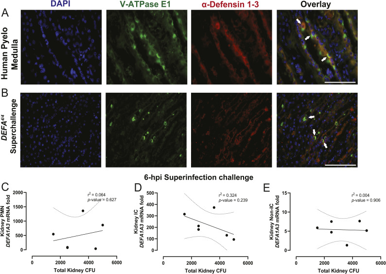
Figure S1. Human and mouse medullary collecting duct–derived intercalated cells co-localize α-Defensin 1-3 expression.
(A, B) Pyelonephritis human– and (B) uropathogenic E. coli–challenged DEFA4/4 mouse kidney sections were stained with V-ATPase E1 (green), α-Defensin 1-3 (red), and nuclear DAPI (blue) markers. Arrows denote co-location events from α-Defensin 1-3–expressing collecting duct epithelial cells (suspected intercalated cells). The white bar indicates 100 μm size for the image sections recorded under a 60X objective lens. (C, D, E) Correlation analysis with indicative Pearson’s coefficients and P-values from uropathogenic E. coli–challenged total kidney bacterial CFUs from isolated (C) leukocytes, (D) intercalated cells, and (E) non-intercalated cells at 6 hpi. The straight line denotes the linear regression with 95 confidence intervals represented with dotted lines for correlation graphs.

