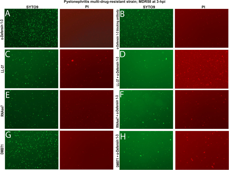
Figure S4. α-Defensin 1-3 peptide bacterial damage and agglutination are enhanced in combination with LL-37, RNase7, and DMBT1 antimicrobial peptides (AMPs) against pyelonephritis multi-drug–resistant E. coli strain in vitro.
(A, B, C, E, G) Visual agglutination of all bacteria (left) and damaged (right) pyelonephritis multi-drug–resistant strain; MDR58 co-incubated with (A) 10 μg/ml α-Defensin 1-3, (B) anti-α-Defensin 1-3 blocking antibody, or 30 μg/ml of each AMP alone, (C) LL-37, (E) RNase7, and (G) DMBT1 peptides did not induce bacterial damaging effects as lacking PI staining. (D, F, H) Antimicrobial effects were visually enhanced when 10 μg/ml α-Defensin 1-3 was co-incubated with each AMP combination. (D) Combination of (D) α-Defensin 1-3 with either LL-37, RNase7, or DMBT1 peptides produced higher density of damaged bacterial aggregates at 3 h post-incubation than when AMP used alone. Images from staining were recorded under a 20X fluorescent lens.

