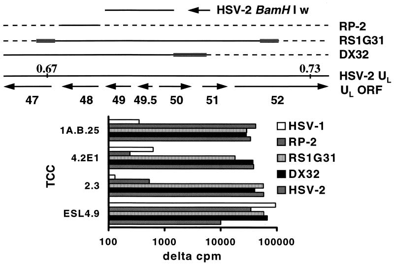
FIG. 1.
(Top) Organization of the HSV genome in the region of map units 0.67 to 0.73. Boundaries are approximate. HSV-1 × HSV-2 IRV are also shown. HSV-2 DNA is indicated by a solid line, HSV-1 DNA by a dashed line, and indeterminate regions by multiple lines. The HSV-2 BamHI w fragment used for expression cloning is also shown. ORF, open reading frame. (Bottom) Proliferative responses of TCC to the indicated IRV. Data are Δcpm, expressing [3H]thymidine incorporation compared to that in the medium, which was less than 500 cpm in each case.

