Figure 4.
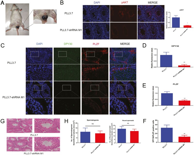
DPY30 inhibition in testis could not induce testicular dysgenesis in mouse. A. The injection of DPY30 interference lentivirus in mice testis. B. Testes were treated with lentivirus for 35 d; the expression of pAKT in testes was analyzed. C. Testes were treated with lentivirus for 35 d; the expression of DPY30 and PLZF was analyzed in the testes. D. The quantitative analysis of DPY30 expression in C. E. The quantitative analysis of PLZF expression in C. F. The DPY30/PLZF positive cell number in C. G. Testes were treated with lentivirus for 35 d; the effect of DPY30 interference lentivirus on mice testicular morphology; H. Testes were treated with lentivirus for 35 d; the effect of DPY30 interference lentivirus on cell counts (spermatogonia, round spermatid cell) in mice testis were calculated. At least three biological replicate samples (mice) for immunohistochemistry and immunofluorescence were analyzed. (B-H) Data were presented as the means ± standard deviation (B-H). Data were compared by two-tailed Student’s t test, *P < 0.05, **P < 0.01, ***P < 0.001 (B-H).

