
FIG. 2.
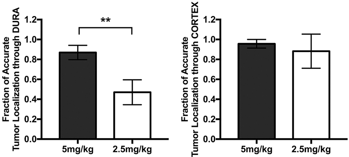
Fractions of tumor accurately localized through the dura and cortex. Left: NIR signal on dura view was evaluated for its ability to guide localization of the tumor. The localization was deemed successful by the independent reviewer if the location of the tumor determined on the dura view corresponded with the location shown on the tumor view. A statistically significant difference in the fraction of accurate tumor localization through dura between the two dose groups was demonstrated (**p = 0.0047). Bars represent the standard error. Right: Postdurotomy, a similar analysis was performed on the cortex view. The difference between the two groups was not statistically significant (p = 0.39). Bars represent the standard error.