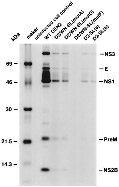
FIG. 9.
Viral proteins in infected LLC-MK2 cells. Viruses derived by transfection were passaged in C6/36 or in LLC-MK2 cells, as described in the legend to Fig. 6, and then used to infect LLC-MK2 cell monolayers at an MOI of 0.05, in parallel with cells used in the analysis of viral RNA synthesis shown in Fig. 7. After 2 days, cells were radiolabeled for 4 h with [35S]promix (methionine plus cysteine). Then cell monolayers were disrupted by trypsinization, and cells were lysed in radioimmunoprecipitation assay buffer. Proteins in the lysate from one 35-mm-diameter well of a six-well plate were immunoprecipitated with DEN2 HMAF, and precipitates were collected with Pansorbin beads. Radiolabeled proteins were analyzed on a tricine-buffered SDS–12% polyacrylamide gel. DEN2 proteins were presumptively identified by size.

