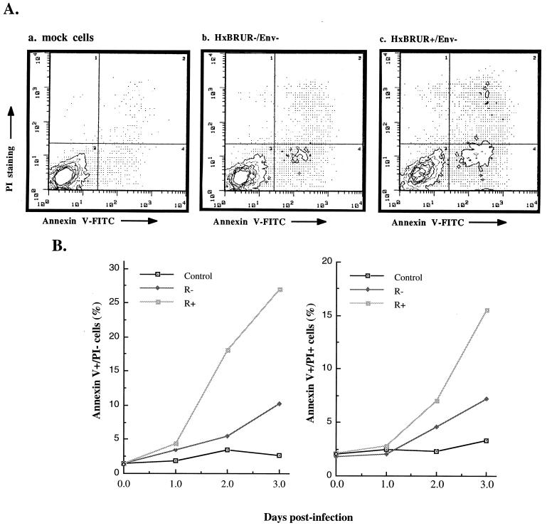
FIG. 2.
Vpr induces apoptosis during single-cycle infection of dividing Jurkat T cells. (A) Jurkat T cells were infected with VSV-G pseudotyped Vpr+ or Vpr− HIV-1 viruses. At 48 h p.i., the infected or mock-infected cells were costained with Annexin V and PI and analyzed by flow cytometry. The axes represent the cell-associated fluorescence intensity of Annexin V (x axis) and PI (y axis). (B) The percentage of Annexin-V+ PI+ or Annexin-V+ PI− cells within the different infected cell populations were evaluated by Annexin V-PI costaining and flow cytometry analysis at 1, 2, and 3 days p.i.

